Critter of the Month is highlighting the arrival of spring underwater this month with a whole community of organisms visible under a microscope this time of year. As on land, spring underwater is marked by a flush of new green growth, here in the form of phytoplankton. Many diatoms (a type of phytoplankton) start to appear in high numbers even in the winter, but many phytoplankton groups grow faster and reach higher biomass as spring temperatures rise, and especially as daylengths grow longer.
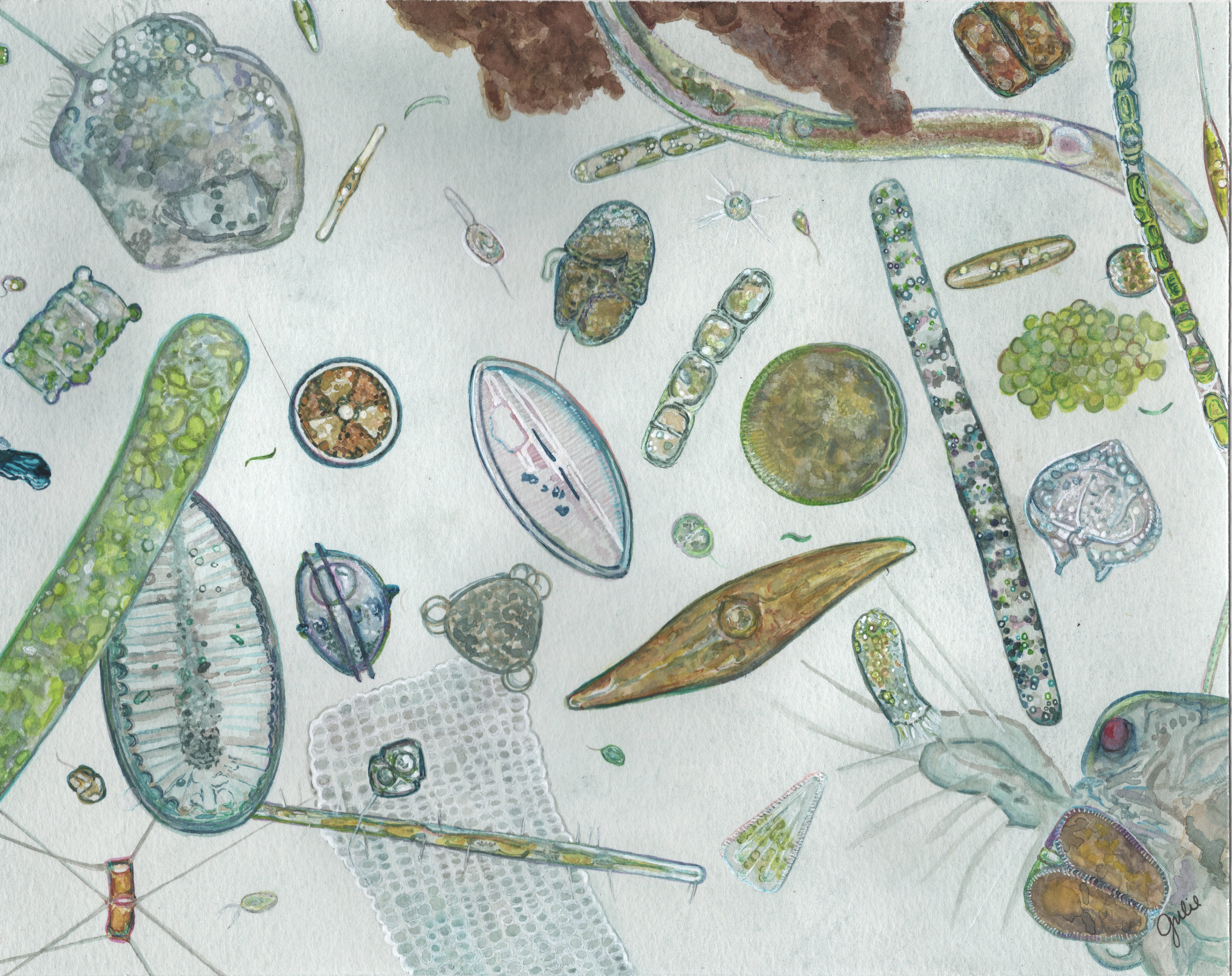
How many organisms can you identify in the image?
Take a close look at the watercolor, and then see our labelled version.
The water color captures a snapshot of the Chesapeake spring bloom. You’re looking at a collection of microbes gathered from a net towed along our research pier. The net has a mesh size of 20 microns, which is quite fine so we can capture some fairly small phytoplankton. But you don't need highly specialized equipment to try this yourself: towing a pair of pantyhose or even a cloth scarf through the water can collect some plankton and give you a first glimpse of what's out there. As you can see in the labelled image, we can identify some organisms with confidence to the genus level, while others we can only place in a broader category. Microscopy remains a gold standard tool for tracking which phytoplankton are present and when. But like any single approach, it has its limits. Even trained eyes, looking at cells that may vary in appearance, or that cannot be reliably distinguished based on appearance alone, can only go so far.
We use multiple tools at the PhytoChop Observatory to identify who's in the water. One key tool, DNA barcoding, lets us detect organisms by their genetic signatures. This approach too has some important limitations and will be the subject of a future essay.
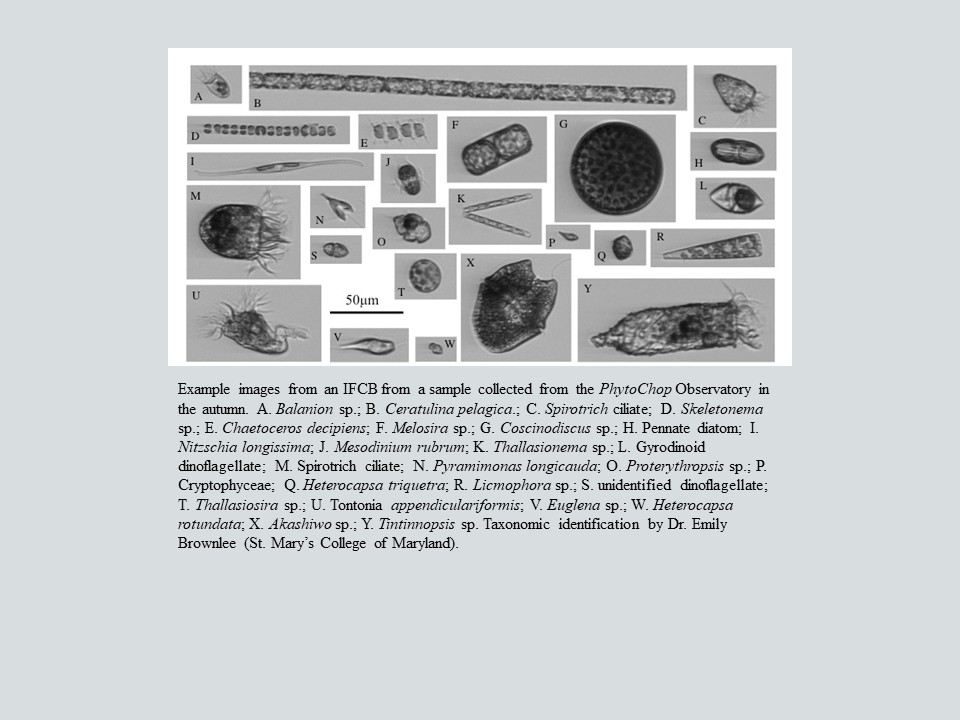
Another key instrument is the Imaging FlowCytobot. The Cytobot continuously pulls Choptank water through a flow cell just 150 microns wide. This is sufficiently narrow that it forces cells to pass through in single file. Living phytoplankton cells are fluorescent due to their chlorophyll, and any fluorescent cell passing through the flow cell between 5 and 150 microns triggers a camera, and an image is captured and stored. This process can result is tens of thousands of individual cell portraits in a single hour. Each image is run through an AI image classifier that assigns an identity based on information it has learned from a training dataset.
The AI image classifier is only as good as its training data, which consists of images that have been annotated by taxonomic experts and typically captured across a wide range of orientations. Building and refining that library with taxonomic expertise is an ongoing effort involving members of the PhytoChop Observatory team and collaborators. One advantage of the Cytobot's continuous recording is that we have a growing image archive. As our training datasets improve, we can go back and re-analyze archived images with better-trained classifiers. An important goal we're working toward is real-time identification, which would enable us to know who is blooming and when. Spring is a very exciting time to be watching!
Looking for More Information?
Imaging Flow Cytobots: https://mclanelabs.com/imaging-flowcytobot/